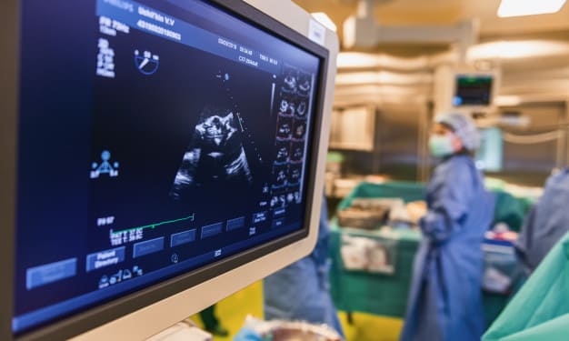
Ultrasound imaging (sonography) is a diagnostic medical procedure that uses high-frequency sound waves to produce dynamic visual images of organs, tissues or blood flow inside the body. The sound waves are transmitted to the area to be examined and the returning echoes are captured to provide the physician with a ‘live’ image of the area.
Let’s discuss some of the innovations in ultrasound imaging that have been introduced in recent areas and also some new technology which is still at the research and development stage.
1. Digital Beam Forming: It improves lateral resolution by accurately focusing the ultrasound beam. Earlier analog beam formers lacked producing delay timers with accuracy but digital beam formers not only produces narrower beams but can also operate in higher frequencies.
2. High-Frequency Imaging: This technology uses frequencies above the normal diagnostic range (above 20 MHz) to produce very high resolution ultrasound images of superficial structures.
3. Extended Filed of View Imaging: This technique enables the operator to produce a static image of a large section.
4. Compound Imaging: This technique improves contrast resolution by scanning from multiple view angles.
5. Three-Dimensional Imaging: This technique requires a volume of tissue to be scanned. There are three approaches to scanning volume of tissue: free-hand, mechanical, and electronic scanning. With the electronic scanning it is possible to produce real time 3-D images.
6. Harmonic Imaging: This technique reduces haze and scatter when scanning large patients. This is achieved by using only a second harmonic frequency echoed to produce the image.
7. Contrast Agents: The main contrast agent used in ultrasound imaging is a solution containing micro-bubbles of air or inert gas. These microbubbles are of a similar size to red blood cells, which enables them to cross the lungs. This enables them to be used for arterial studies using a venous injection.
8. Pulse Inversion Imaging: This imaging modality is used to increase the sensitivity of ultrasound to contrast agents. It improves the axial resolution as the pulse length is shortened.
9. Elastography: This technique is under development, which attempts to distinguish malignant tissue from normal tissue by measuring the amount that a structure distorts under pressure. Ultrasound imaging is used to make the measurement.
10. Tissue Characterization: This technology aims to analyze the signals received from different tissues and characterize them according to their acoustic properties but is still in research stage.
11. Tissue Motion: This imaging is a development of color Doppler and is used to look at the movement of tissues within body organs such as the heart.
12. Portable Ultrasound Machines: These machines have undergone major improvements over the last 10 years and better machines perform as well as conventional scanners.
Are you interested in learning more about these technologies? AIHT Education is an accredited healthcare training school in Stratford CT that offers Cardiovascular Technologist training.
Visit our website at www.aiht.edu to schedule an appointment to speak with an admission counselor. Our friendly and knowledgeable counselors will help you get acquainted with AIHT and our professors, discuss our training programs and answer any and all questions you may have. You can also read our article on how to become an ultrasound technician.