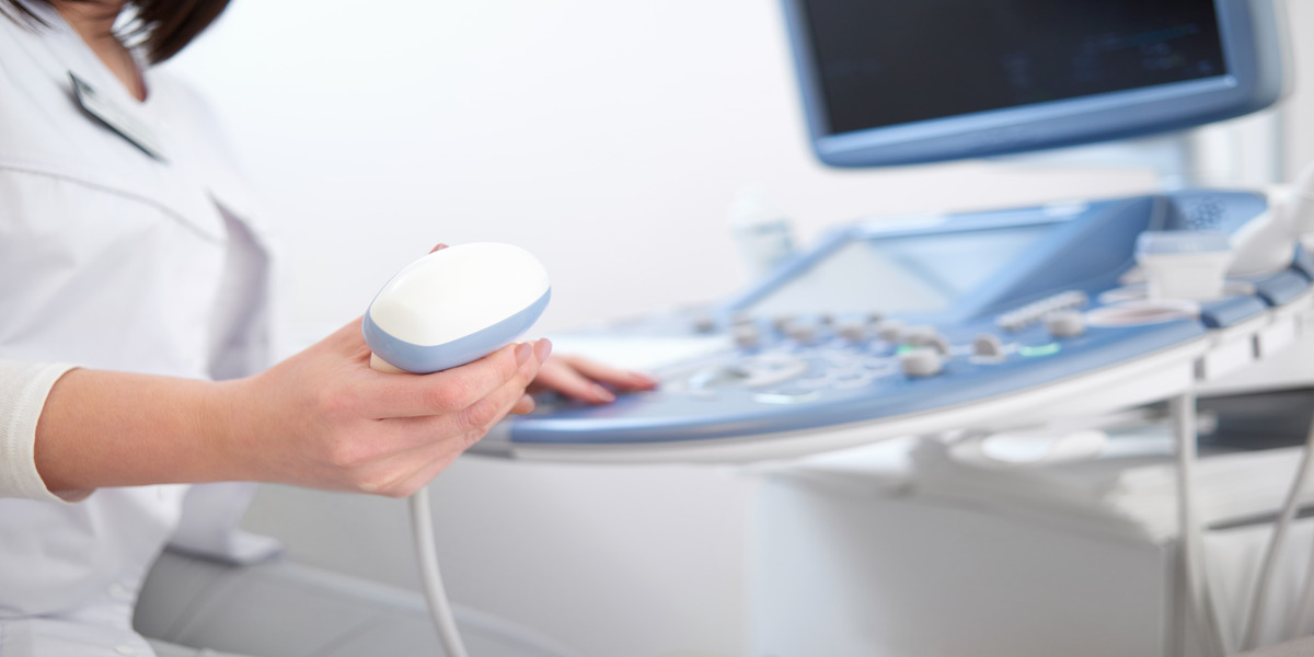
A sonographer is a person who takes ultrasound images of the tissues and organs in the human body. Ultrasound devices are used to accomplish this. Additionally, the heart’s blood vessels and muscles can be photographed by sonographers. Unlike X-ray, sonography does not use radiation. A sonographer can focus on many different parts of the body.
As per the Bureau of Labor Statistics, employment in the career of sonography is estimated to grow 10% by 2031. These statistics shows that if you are planning to make your career in diagnostics medical sonography then it would be great choice!
There are many different types of sonographers and different fields of sonography in which they can specialize, ranging from gynecology and obstetrics to the heart.
What is a Sonographer?
Sonographers are medical professionals who use ultrasonic medical imaging equipment to learn about and diagnose patients’ medical conditions. Some sonographers are also known as ultrasound technicians because they spend a lot of time operating ultrasound equipment.
Different Type Of Sonographers
The following is a list of the several kinds of sonographers, along with some information about their duties:
1. Diagnostic Cardiovascular Sonographer
Sonography is a tool that diagnostic cardiologists use to help doctors diagnose diseases that could harm the heart and cardiovascular system. Cardiac sonographers commonly known as diagnostic cardiovascular sonographers or echocardiographers by some specialists because they use echocardiograms to capture diagnostic images of patients’ internal systems.
Additionally, when taking diagnostic photos to look for abnormalities like blockages or degeneration, diagnostic cardiovascular sonographers frequently examine the heart’s anatomy in both 2D and 3D. After that, doctors can use this information to prescribe treatments and diagnose issues.
However, they can also work in clinics or doctor’s offices with on-staff heart specialists, and diagnostic cardiovascular sonographers typically find employment in hospitals.
2. Obstetric Sonographer
Imaging the fetus while it is still in the womb is the area of expertise of an obstetric sonographer. They look for abnormalities or pregnancy complications besides average growth and development. The sonographer’s job is to ensure the mother and fetus are healthy.
The technician confirms the woman’s pregnancy, examines the fetal position, and can assist in determining the due date. In their diagnostic medical sonography education, a technician will receive instruction in this specialized field because obstetric sonography is one of the most common types of ultrasounds.
3. Abdominal Diagnostic Medical Sonographer
Using sonography equipment, abdominal diagnostic medical sonographers take images of the internal organs in the abdomen. Abdominal sonographers work in various medical settings, including hospitals, clinics, and other facilities that treat and cure patients with conditions related to abdominal regions.
Most abdominal sonographers receive extensive training in the various abdominal systems to effectively assist physicians in diagnosing medical conditions within them because they focus specifically on the abdomen. Abdominal sonographers can look for abnormalities such as tumors, tissue damage, or stones while taking images of patients’ abdominal areas.
Read More:- Reasons You Should Become a Diagnostic Medical Sonographer
4. Musculoskeletal Sonographer
This sonographer takes images of the muscles and other parts of the skeletal system to make a diagnosis. The patient’s tendons, ligaments, joints, and nerves are all affected by this. Indeed, diseases that impair a patient’s mobility by affecting their joints, muscles, and bones are frequently identified by sonographers who specialize in the musculoskeletal system through the images they capture.
Additionally, musculoskeletal sonographers often work in healthcare facilities that treat patients with traumatic injuries, such as emergency rooms and hospitals. During their work, musculoskeletal sonographers keep an eye out for the following conditions: sprains, tense muscles or objects, arthritis, hernias, cysts, bone fractures, and inflammation.
5. Neurosonology Sonographer
A Neurosonology sonographer with adequate ultrasound training uses specialized tools to capture diagnostic brain images in addition to ultrasound and sonogram technology. The transcranial doppler (TCD) machine is the instrument that Neurosonology sonographers use to obtain inward pictures of the mind. It helps doctors diagnose conditions like Down syndrome, encephalitis, and cerebral palsy.
Neurosonologists also use a TCD machine to take pictures of patients’ spinal columns and neurological systems to look for anomalies like strokes, epilepsy, aneurysms, and brain tumors. Due to the specific requirements of their field, most Neurosonology sonographers use TCD machines in hospitals and diagnostic testing facilities.
Read More:- 4 Skills that Make a Great Sonographer
6. Breast Sonographer
After a patient has an abnormal mammogram or examination, a breast sonographer is specialized in taking diagnostic images of the breast and the surrounding tissue. Imaging the breasts, tissue, and lymph nodes is typically part of their job to look for signs of a developing medical condition.
Tumors, cysts, and lumps are some abnormalities breast sonographers frequently look for in patients. Because the images that they take can show areas that might indicate the potential for cancerous growth, breast sonographers can also assist specialists and doctors in diagnosing cancer.
Breast sonographers typically work in hospitals, oncology centers, or women’s health centers due to the primary focus of their work on breast imaging.
An Overview of Sonography Technology
In many instances, a sonogram is finished in a flash. You can anticipate the following:
Prior to the test
Healthcare providers typically order sonography as a first-line test alongside blood tests. Before your sonogram, make sure you ask your provider if you need to follow any special instructions. In a crisis setting, sonography will regularly be performed immediately. Find out if you should or shouldn’t eat or drink before a test that will take place in the future. The purpose of the test may influence the answer.
Read More:- How Can I Become An Ultrasound Technician?
All through the Test
A solitary professional right leads an ultrasound image at the bedside. You will be asked to lie down on the bed and undress sufficiently to expose the test area by the technician. The technician will apply conductive gel to the transducer, which has the consistency of lubricant jelly. The gel will be warm, depending on the available tools and supplies. The technician will then use firm pressure to slide the transducer over the skin. The pressure may occasionally cause slight discomfort.
The technician will use the computer to take pictures and may use a mouse to drag lines across the screen while using the transducer to point to areas of interest. Like a virtual yardstick, the lines assist in measuring size. You ought to be able to observe the entire procedure and even ask questions during it.
Post-Test
Typically, the technician will provide a towel to remove the conductive gel after the sonogram. You will be permitted to change into your clothes once the technician confirms that all necessary images have been taken. There are no additional instructions or side effects to deal with.
Primary duties of a Sonographer
Sonographers need to be in good health to do their jobs. Sonographers ought to be able to carry out the typical tasks listed below in addition to knowing how to use the transducer and other equipment:
- Sonographers must regularly lift 50 pounds
- Stoop and bend
- Have a full range of motion in their shoulders, hands, and wrists
- Be able to hear clearly to distinguish sounds from monograms
- They must also be able to stand for long periods
- Assist patients in getting on and off examination tables
- Complete the sonogram in the correct order
Key Skills Required for Sonographers
Sonographers can be skilled in the following areas and domains:
1. Physical ability
The occupation of a sonographer requires a few physical capacities. A lot of sonographers spend a lot of their workday standing up. During an imaging session, they may lift or position patients, so it is essential to have the strength and endurance to move patients. For some imaging methods, sonographers also need to have good hand-eye coordination.
2. Technical proficiency
Sonographers are able to make efficient use of a variety of specialized medical imaging tools. Accurate medical images are essential for diagnosing abnormalities, evaluating options for treatment, and tracking the healing process of illnesses and injuries.
As a result, it is necessary for sonographers to have thorough training in the medical equipment they use in order to guarantee that patients receive essential care.
3. Attention to detail
Medical images are created and evaluated by sonographers. Accurate pictures of internal body structures are taken with the appropriate tools. They accurately label, organize, and file the images they produce so that they are simple to access and identify.
Sonographers and physicians sometimes collaborate to analyze the imaging results. They use their keen eye for detail to spot unhealthy tissues, minor bone fractures, and abnormalities in the development of tissue or bone.
4. Communication
Sonographers need to be able to communicate effectively because they spend a lot of their workday interacting with patients and other members of the health care team.
These team members may include additional sonographers, physicians, nurses, insurance providers, technicians, and other medical specialists, depending on the setting in which they work.
Sonographers use their communication skills to adapt their style to meet the requirements of various audiences when working with these diverse groups. Additionally, they must hold a current certification required for the diagnostic medical sonographer.
FAQ
Q1: What’s the difference between sonography and ultrasound?
Similar to asking about the difference between a carpenter and a hammer, asking about the difference between sonography and ultrasound is identical. This is since, just as a carpenter uses a hammer for a specific task, a sonographer uses ultrasound equipment for a variety of tasks. Sonography is a medical skill; the device is ultrasound.
- The Science of Sonography
- Ultrasound – The Images
Sonographers acquire knowledge of the science of sonography, which literally translates to “sound writing.” Because sonographers use ultrasound or high-frequency sound, they create images. This is why the terminology “ultrasound writing” is utilized to define it. Most of the words that start with ultra- are fun to say. For instance, ultracrepidarians are people who criticize or judge outside of their field of expertise; that is, they are ultrarapid (no, they really do).
Ultrasound machines produce and receive ultrasounds, which are sound waves with a very high pitch. The highly skilled ultrasound technologist makes use of ultrasound machines to assist medical professionals in “seeing” inside patients. Today’s ultrasound equipment can produce stunning images. Pictures in two dimensions and three dimensions are both possible, and four-dimensional movies can be made with a bit of computer magic.
Q2: What are the main types of sonographers?
The most common types of sonographers include diagnostic medical sonographers, vascular technologists, and cardiac sonographers. Diagnostic medical sonographers specialize in imaging the organs and tissues of the abdomen, pelvis, and obstetric and gynecologic areas. Vascular technologists focus on imaging the blood vessels, including those in the neck, arms, legs, and abdomen. Cardiac sonographers specialize in imaging the heart and its blood vessels.
Other types of sonographers include pediatric sonographers, who specialize in imaging children and infants, and musculoskeletal sonographers, who specialize in imaging the muscles, tendons, ligaments, and joints of the body. Additionally, some sonographers specialize in performing ultrasound-guided procedures, such as biopsies or aspirations.
Each type of sonographer requires specialized training and certification. If you are interested in pursuing a career as a sonographer, it’s important to research the different types of sonography and choose a specialty that interests you.
Q3: What are the 3 types of ultrasounds?
The standard sorts of ultrasounds are:
- Abdominal ultrasound, which examines the liver, gallbladder, pancreas, and spleen, among other internal organs in the abdomen.
- Obstetric/pregnancy ultrasound is a routine test to check on the baby’s health and growth. This also includes female pelvis ultrasound, which can examine the female pelvis, uterus, cervix, fallopian tubes, and ovaries using external pelvic ultrasound or transvaginal ultrasound (with the transducer in the vagina).
- Transrectal ultrasound, which provides images of the prostate gland, and renal ultrasound, which is used to scan the urinary tract, including the kidneys and bladder.
Q4: What is the highest level of sonography?
Professionals in ultrasound technology view internal organs on a computer monitor by using sound waves from an ultrasound machine. There are associate, bachelor, and master’s degree programs in ultrasound technology. Regardless of degree level, a program in ultrasound technology ought to be accredited and enable graduates to take the credentialing or registration exam for their profession.
Q5: Is sonography in high demand?
Indeed, clinical sonographers are sought after the nation over. Medical sonographers with the necessary skills are needed to join the care teams at numerous healthcare facilities, including clinics, doctor’s offices, and hospitals. The U.S. Bureau of Labor Statistics predicts that demand will only continue to rise. Diagnostic medical sonographers’ growth rate is much faster than the average, at 14%, compared to the 8% growth rate for all professions.
Q6: How to become a diagnostic medical sonographer?
There is a high demand for sonographers, and starting pay is excellent. The vocation resembles many high-level care professions. Your job is skilled, stimulating, and varied, and it provides valuable patient care information. A two-year degree from an accredited sonography training program is the most common educational path for prospective sonographers. Bachelor’s degrees and one-year certificate programs in sonography are also available for individuals with prior training in another healthcare field.


